SINAPSE Early Career Researcher Exchange Fund 2022
This SINAPSE funded programme sent PhDs and ECRs to work with companies and overseas research labs to give them greater experience of techniques and developments they could bring back to use in their research.
Creation of CE-MMUS tissue phantoms and development of magnetic phase-change nanodroplets
Funding Recipient: Georgia Adam, Biomedical Engineering, University of Strathclyde
Supervisors: Dr Helen Mulvana, Biomedical Engineering, University of Strathclyde
Prof Carmel Moran, Queens Medical Research Institute, University of Edinburgh
Prof Tomas Jansson, Biomedical Engineering, Lund University
Partner Organisation: University of Lund, Sweden
Project Report: My current work is focussed on a novel ultrasound based imaging modality which involves combing two forms of contrast agents, superparamagnetic iron oxide nanoparticles (SPIONS) and microbubbles (MBs), to ultimately improve early detection of lymph node metastasis in colorectal cancer. Before any experimental work was carried out, simulation studies indicated that the complex of SPION-MBs could yield larger tissue displacements, improving sensitivity to metastatic stiffness [1]. This research exchange was focussed on two main objectives for progression from simulation studies to experimental work for validation purposes. The first objective involved the development of a tissue mimicking material (TMM) which is able to accommodate the embedment of microbubbles, one of our contast agents, in its matrix for direct comparison between Magnetomotive Ultrasound (SuperParamagnetic Iron Oxide Nanoparticles (SPIONS) alone) and Contrast Enhanced Magnetomotive Ultrasound (Microbbubles (MBs) conjugated to SPIONS through a streptavidin-biotin bond) for increasing the imaging sensitivity by yielding higher tissue displacement values when subjected to the same excitation frequency. The second objective was focussed on a technique for determing the maximum loading of SPIONs that could be achieved per microbubble by using Nuclear Magnetic Resonance (NMR) to ensure complete conjugation of all the SPIONS to the Microbubbles before fabrication and incorporation in the TMM material. Our initial discussions with the fellow institution was focussed on which material may be appropriate for the synthesis of our tissue mimicking material (TMM). Our best candidate was a polyacriolimide hydrogel material (PAAm) for which stiffness can be varied by only increasing the water content during polymerisation. Fabrication protocol of this material is found in [2]. According to stiffness values for healthy lymph nodes based on [3] inguinal lymph node was measured to be about 10 kPa. For the values of the stiffness we are interested in, an amount of 20g of water was used for the fabrication of the PAAm. Initially the hydrogel was made using the 4 components (no SPIONS included). Next and according to [4] SPIONS were added in the solution corresponding to 0.6mg of SPIONS/ml. It needs to be mentioned at this stage only SPION loaded PAAm samples were fabricated before carrtying out the work needed to ensure complete conjugation of SPIONS to MBs as desceribed next.
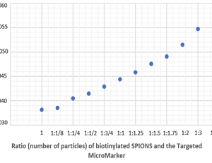
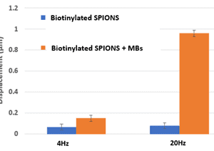
1H Nuclear Magnetic Resonance (NMR) measurements using an Agilent/Varian VNMR-S 500MHz was used to investigate any alterations on the magnetic properties of SPIONS (more specifically biotinylated SPIONs (SPIONS-B) to be able to bond with the streptavidin coated MBs) and increasing ratio of MBs. 1H NMR measurements of SPIONs-B to increasing TR-MM ratio was used to determine maximum loading of SPIONs that could be achieved per microbubble. For these measurements the T2 relaxation time of the proton peak was used for each dilution ratio. Data showed a gradual narrowing of the proton peak (increasing T2) to the point saturation occurs at a ratio of 1:3, indicating SPIONs-B have less surface area exposed to water due to complete binding to TR-MM. Knowing the ratio of these two contrast agents was vital to ensure complete conjugation of all the SPIONS to the MM before fabrication and incorporation in the TMM material. After determining the ratio of rhe SPIONS to MBs the PAAm hydrogel was fabricated and examined. Preliminary data of displacement values for the PAAm containing SPIONs-B or SPIONs-B + MBs. These experiments were carried out using a in house build solenoid (0.14T, 4Hz, 20Hz sin) and imaged with VEVO 3100. It can be noted that SPIONs-B + MBs display a trend towards higher displacement especially for the 20Hz case initially verifying the silulation findings.
Overall the funded by SINAPSE Knowledge Exchange grant yielded important results which are currently being used for preparing a peer reviewed journal publication. The successful development of a TMM for use with both contrast agents to directly compare tissue displacement values and the novel way of determining the loading of SPIONs on the MBs using NMR is hoping to have broader appeal across SINAPSE and possible use from groups carrying out similar work.
[1] Sjöstrand S, Bacou M, Kaczmarek K, Evertsson M, Svensson IK, Thomson AJ, Farrington SM, Moug SJ, Jansson T, Moran CM, Mulvana H. Modelling of magnetic microbubbles to evaluate contrast enhanced magnetomotive ultrasound in lymph nodes – a pre-clinical study. Br J Radiol. 2022 Jul 1;95(1135):20211128. doi: 10.1259/bjr.20211128. Epub 2022 May 19. PMID: 35522781.
[2] Penkavova, Vera & Spalova, Anna & Tomas, Jan & Tihon, Jaroslav. (2022). Polyacrylamide hydrogels prepared by varying water content during polymerization: Material characterization, reswelling ability, and aging resistance. Polymer Engineering & Science. 62. 10.1002/pen.25895.
[3] Jung JW, Je H, Lee SK, Jang Y, Choi J. Two-Dimensional Shear Wave Elastography of Normal Soft Tissue Organs in Adult Beagle Dogs; Interobserver Agreement and Sources of Variability. Front Bioeng Biotechnol. 2020 Aug 19;8:979. doi: 10.3389/fbioe.2020.00979. PMID: 32974311; PMCID: PMC7466577.
[4] Evertsson M, Cinthio M, Fredriksson S, Olsson F, Persson H, Jansson T. Frequency- and phase-sensitive magnetomotive ultrasound imaging of superparamagnetic iron oxide nanoparticles. IEEE Trans Ultrason Ferroelectr Freq Control. 2013 Mar;60(3):481-91. doi: 10.1109/TUFFC.2013.2591. PMID: 23475915
Mobile Cognition and mobile electroencephalographical (EEG) research
Funding Recipient: Emily Cunningham, Psychology, University of Stirling
Supervisors: Dr Christian Keitel, Psychology, University of Stirling
Prof Stefan Debener, Department of Psychology, University of Oldenburg
Partner Organisation: University of Oldenburg, Germany
Project Report: I am a first-year PhD student at the University of Stirling, and my project is dependent on collecting high-quality EEG data in mobile settings (i.e., outside of the lab). My work focusses on investigating neurophysiological markers of “lapses” in attention, both in laboratory and ‘real-world’ settings, with the overall aim being not only to examine whether we can identify markers of inattention, but moreover whether these markers can be reliably identified outside of the lab. The project requires specialist equipment, and Professor Debener is a co-developer of the mBrainTrain Smarting Pro: a new and innovative mobile EEG system which allows participants free range of movement in various settings (even including, for example, while swimming). The purpose of my visit was therefore to gain specialist knowledge with using this system and conducting mobile EEG research from world-leading researchers, and co-developers and experts in the specific equipment that I will be using.
The aims and expectations of this visit were met and exceeded, and the benefits were two-fold: I received specialist knowledge of, and experience with the mBrainTrain Smarting Pro EEG system, and I also learned about aspects of processing mobile EEG data, specifically using EEGLAB. Following the successful exchange, my primary supervisor, Dr Christian Keitel, has purchased the mBrainTrain Smarting Pro system and I will be able to begin using it for my own work soon.
The opportunity to do something like this at such an early stage in my career has been extremely beneficial to my development as an independent researcher: it allowed me to reflect on my training needs and ensure these were met during the exchange, discuss my research plans with others, and begin to build connections internationally.
Mobile Cognition is a new field of research and collaboration between researchers is crucial. This exchange has bridged connections between the University of Stirling (Scotland) and the University of Oldenburg (Germany): an institution renowned for its mobile EEG research. The knowledge gained from this exchange will be invaluable for designing and conducting our research here in Stirling, which will then be disseminated via publications, conferences, teaching, and visits to other institutions, allowing for research in the field of Mobile Cognition to expand and continue to develop.
Deep neural networks (DNNs) in OCT-angiography (OCTA)
Funding Recipient: Jinpeng Liao, Biomedical Engineering, University of Dundee
Supervisors: Prof Zhihong Huang, Biomedical Engineering, University of Dundee
Prof Ruikang Wang, Biomedical Engineering, University of Washington
Partner Organisation: University of Washington, Seattle, USA
Project Report: This exchange project was supervised by Ruikang Wang, a professor of bioengineering and ophthalmology at the University of Washington. He is the Director of Biophotonics and Imaging Laboratory. During the study, I visited sixteen state-of-the-art optical coherence tomography (OCT) imaging systems, including swept-source OCT (SSOCT), spectral-domain OCT (SDOCT), and polarization-sensitivity OCT (PSOCT) in his laboratory. Among these systems, I focused on understanding the system configuration of SSOCT, and the development of algorithms for OCT image processing and angiography signal extraction based on repeated OCT scans. Furthermore, with the abundant retina OCT data in his lab, I investigated a deep-neural network for OCT image reconstruction.
Due to the current limitations of OCTA imaging in the retina application, I investigated a series of methods to reconstruct the quality of OCTA image, including a Frangi filter for vessel enhancement, a deep neural network (DNN) for image reconstruction, and an improvement of the OCTA generation algorithm. Furthermore, during the exchange, I learned the clinical setup of the hand-held SSOCT system and understood the clinical OCTA retina scanning workflow.
In terms of the extensional function of OCTA imaging, I studied algorithms to detect capillary velocimetry, with the support of the laboratory at the University of Washington.
Jinpeng has been invited to return to the UoW lab for a full year, leaving in April 2023.
quantitative Magnetic Resonance Imaging (qMRI) in rapid acquisition of diffusion imaging in relation to pathological processes following exposure to football heading
Funding Recipient: Ali Muqtadir, Psychology, University of Stirling
Supervisor: Dr Magdalena Ietswaart, Psychology, University of Strathclyde
Partner Organisation: Institute for Neuroscience and Medicine (INM-4), Jülich, Germany
Project Report: I am a first-year PhD student at the University of Stirling working on a multi-methods approach to investigating the effects of football heading on brain health, supervised by Dr Magdalena Ietswaart. The goal of my research exchange was to collaborate with the Medical Imaging Physics department at Research Center Julich, Julich, Germany to take advantage of their expertise in 7T sequence development and MR methods and learn how they can be put to use in investigating a clinical issue.
I travelled to Germany to work with and learn from the research groups at RWTH Aachen and the Institute of Neuroscience and Medicine of Forschungszentrum Jülich who are collaborating with Stirling on piloting a multi-method study of the effects of football heading with a comprehensive battery of measures including EEG, blood sampling, and most notably 7 Tesla MR scanning. With researchers from Aachen, I acquainted myself with the study protocol and underwent all measures myself to better understand the participants’ experience of it. This included becoming competent in the use of the EEG system and the proper handling of blood samples. The majority of my time in Germany was spent working with colleagues at Julich, where the 7T scanner is located. The goal of my time in Julich was to familiarise myself with the MR sequences being used in the study and get a start on learning to analyse them. During my time there I received instruction in handling data coming from the 7T scanner as well as analysis of Magnetic Resonance Spectroscopy, structural MP2RAGE, and Susceptibility Weighted Imaging data. The high resolution of the 7T scanner results in unparalleled detail compared to scanners with lower field strengths, however, the stronger signal comes with increased noise which poses an issue for traditional MR analysis pipelines. In my time at Julich, I worked closely with medical physicists on trying to come up with the best way to segment and coregister images coming from the 7T scanner while filtering out the noise. In addition to learning about how MR data is collected and analysed, I had the chance to experience first-hand what it’s like to be in a 7T scanner. As it was my first time in an MRI machine, this gave me great insight into what the patient/participant experience is inside the scanner.
The Medical Imaging Physics department at Julich is renowned for MR sequence development. With our focus on using quantitative MRI measures in looking at heading impacts, the researchers at Julich have been the ideal collaborators. Their expertise in developing fast sequences for water mapping have meant that we can use more sequences in our imaging protocol to get a more comprehensive picture of what happens to the brain immediately after heading. Learning about these 7T sequences was a highlight of the exchange and I am excited to bring that knowledge back to Scotland and apply it to future projects as well.
Thanks to this research exchange I have become more familiar with key methodologies including MR methods and EEG that are going to play a central role in my project going forward. In addition to the methodological skills that I have been exposed to during the first three months of my studentship, the one aspect of my time in Stirling and Germany that has proven to be most rewarding has been connecting with the research community in both locations. The expansive nature of this project necessitates collaboration across disciplines. The ideas and instruction from established researchers, as well as fellow students, have been invaluable for me in not only getting up to speed on the project but also adjusting to life as a PhD student.
Cross-validating MRI methods to probe tissue microstructure in multiple sclerosis (MS)
Funding Recipient: Dr Elizabeth York, University of Edinburgh
Supervisor: Professor Adam Waldman, University of Edinburgh
Partner Organisation: Cardiff University Brain Imaging Research Centre (CUBRIC), Cardiff University, Wales
Project Report: At the end of 2022, I was fortunate enough to be awarded SINAPSE Early Career Research Exchange (SINAPSE22) funding to visit Cardiff University Brain Research Imaging Centre (CUBRIC). CUBRIC is home to a CONNECTOM high-gradient MRI system, the only one of its kind in the UK and one of only six systems worldwide, which permits imaging with higher signal-to-noise, high b-values and shorter echo times; this has particular advantages for axon diameter distribution mapping, microstructural parameter estimation in vivo. The aims of my exchange visit were to enable cross-validation of MRI methods to probe tissue microstructure, including diffusion-weighted neurite density imaging (e.g. NODDI), magnetization transfer imaging (e.g. MTsat) and aggregate measures (e.g. g-ratio) for clinical translation to multiple sclerosis.
Over two visits, we performed a ‘travelling head’ experiment in which MR images, including diffusion-weighted imaging, were acquired on (1) CONNECTOM and (2) Prisma Siemens MR systems at CUBRIC, Cardiff and (2) Prisma Siemens MR system at the RIE, Edinburgh. Image acquisitions on Prisma systems at the two sites were identical and mirrored the FutureMS MR acquisition protocol (see Meijboom et al. (2022) Wellcome Open Research), plus an additional HARDI (High-Angular Resolution Diffusion Imaging) acquisition. Acquisition protocols on the Connectom included a HARDI acquisition and high b-value (b=30,000) “powerlaw” acquisition for axonal diameter mapping.
I also had the opportunity to hear about ongoing research at CUBRIC from Professor Derek Jones and many other brilliant minds, and discuss potential future collaborative projects. CUBRIC researchers kindly gave up their time to explain their work on topics such low-field hyperfine MRI, TMS/EEG, the WAND cohort, Soma and Neurite Density Imaging (SANDI), machine learning, and sodium imaging. This stimulated further discussions and hypotheses. Of particular interest were initial discussions with Professor Mara Cercignani and neurologist Dr Emma Tallantyre centred around collaborative funding applications for research on multiple sclerosis (MS). Since my initial exchange visit, these discussions have continued and plans for collaborative projects building on joint expertise in microstructural imaging and clinical studies of MS.
One of the key pieces of work that has come out of the exchange visit is development of methods to transfer image quality from high-performance scanners such as the connectom to images acquired in a clinical setting for conditions such as MS. Previous research has developed tools for transferring image quality (e.g. the Image Quality Transfer toolbox https://github.com/ucl-mig/iqt) but further work is needed to make these methods applicable to images acquired in clinic, where pathology may disrupt these algorithms. In addition, we hope the powerlaw acquisition may help to better inform estimation of the g-ratio by giving a better approximation of axonal diameter than lower b-value acquisitions.
Although I already had gained some knowledge of application of microstructural MRI methods in MS during my PhD and post-doctoral work at University of Edinburgh, the exchange visit to CUBRIC was enormously beneficial. In particular, it allowed me to experience first-hand microstructural acquisition protocols which are not possible on systems currently available in Scotland. The non-clinical setting also allowed some degree of “experimentation” which a clinical setting may not readily allow due to resource allocation. Secondly, researchers at CUBRIC have a wealth of experience in microstructural imaging, particularly diffusion-weighted imaging (DWI), and have well-developed pipelines for processing these images. This enabled knowledge exchange of different acquisition and analysis methods.
In the long-term, this exchange has provided me with a valuable opportunity to develop further understanding and transferable skills in advanced microstructural neuroimaging. My visit has begun to foster collaborations between the Cardiff and Edinburgh imaging groups that will be supported by targeted joint project funding proposals. Work is currently is still ongoing from initial MRI acquisitions but I hope to use the expertise gained and academic outputs from the exchange to also support applications for personal research fellowships to develop as an independent researcher. I would like to thank SINAPSE, Professor Derek Jones and all the researchers at CUBRIC who enabled my visit and made it such a success.