SINAPSE Early Career Researcher Exchange Fund 2024
This SINAPSE funded programme sent PhDs and ECRs to work with companies and overseas research labs to give them greater experience of techniques and developments they could bring back to use in their research.
An Insight into Cerebral Amyloid Angiopathy – Zooming in on Cerebral Small Vessel Disease
Funding Recipient: Angelina Kancheva, PhD student Precision Medicine DTP, University of Glasgow & Edinburgh
Supervisors: Professor Terry Quinn, University of Glasgow
Dr Donald Lyall, University of Glasgow
Prof. Joanna Wardlaw, University of Edinburgh
Partner Organisation: Massachusetts General Hospital, Harvard Medical School, Boston MA, USA
Project Report: I am currently a third year PhD student on the Precision Medicine Doctoral Training Program, primarily based at the University of Glasgow but also affiliated with the University of Edinburgh. The main focus of my PhD research is to better characterise the complete clinical phenotype of cerebral small vessel disease (cSVD), a leading cause of stroke and vascular dementia. This year, I get to diversify my PhD skills by undertaking a project focused on plasma biomarkers of Alzheimer’s disease (AD) and their role in predicting cognitive impairment.
Last year, I was fortunate to be one of the recipients of SINAPSE Early Career Researcher Exchange Fund (SECRE). In November, I spent four wonderful weeks at Dr. Susanne van Veluw’s Translational Cerebral Amyloid Angiopathy (CAA) Research Lab based at Massachusetts General Hospital Harvard Medical School. The aim of my research visit was to learn more about CAA, one of the two most common forms of cSVD, the pathology of which also overlaps to a significant extent with that observed in AD.
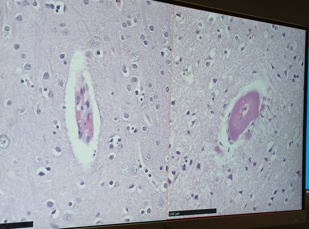
As part of my research exchange, I familiarised myself with some of the methods and techniques used in the lab to unravel the histopathological markers of CAA, which can be used to infer mechanisms involved in the disease. As a clinical researcher mostly used to working with large datasets, I was incredibly excited about being in the lab for the first time. I had the opportunity to shadow a good variety of activities, for example, a two-day double immunofluorescent green fluorescent protein (GFAP), At8 and Thioflavin S staining experiment for the presence of astrocytes (a subtype of glial cells in the human central nervous system, which perform structural, metabolic and neuroprotective tasks), tau and amyloid beta protein (the main proteins involved in AD, which are also found in CAA) in human CAA brain tissue, respectively. It was fascinating to change the perspective through which I investigate and think about cSVD, and glimpse into the workflow of pre-clinical and translational SVD-related work.
Since a lot of the research carried out in Dr. Van Veluw’s lab involves animal work, I also got to shadow an immunohistochemistry experiment with Resorufin and Thioflavin S, which are both dyes that can be used to stain for presence of amyloid protein, on free floating sections of a mouse brain. It was interesting to compare the different experiments that I observed and reflect on the complexity of working with mice in a lab setting. On that note, one of the most intriguing procedures I got to shadow was a mouse craniotomy surgery, which prepared a mouse for two-photon imaging of an area of the visual cortex. I witnessed all steps involved, from animal preparation, anaesthesia delivery, through exposing the region of interest in the mouse brain, to returning the animal to its cage. This was a humbling experience, which helped me appreciate just how much focus, attention to detail, and consideration is required when working with an animal under surgical conditions.
During my time with the Translational CAA group, I was also invited to give a talk at one of the regular J. Philip Kistler Stroke Research Center meetings. My talk entitled “From Cerebral Small Vessel Disease to Alzheimer’s Disease: Insights from Population-Based and Biomarker Studies” was an excellent opportunity to talk about some of the analysis methods I have used as part of my PhD thus far, including phenome-wide association study (PheWas). It was beneficial to meet the wider research group, attended by research fellows, scientists and academics involved in both clinical and preclinical/translational research, as we were able to exchange and discuss ideas, which would be valuable to my research back in Glasgow.
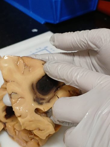
Last but not least, some of the other activities I partook in included a 3T magnetic resonance imaging (MRI) session of a CAA patient at the Athinoula A. Martinos Center for Biomedical Imaging, a world-leading research centre devoted to development and application of advanced biomedical imaging technologies, inspecting a human brain with CAA pathology under supervision, as well as attending several research group meetings and two Neurology lectures in the Ether Dome, the site of the first public demonstration of the use of inhaled ether as a surgical anaesthetic in 1846. All these experiences were enriching and enabled me to appreciate how advanced neuroimaging, immunohistochemistry and immunofluorescence can be used in concert to elucidate disease hallmarks and mechanisms.
In addition to learning about the research that the group is involved in, I found it stimulating and inspiring to spend a month living in Boston, the ‘thinking centre’ of the United States, a city that has some of the most educated people in the world condensed into one area, and one that is rich in history and culture. I was able to enjoy Harvard campus and visit Massachusetts Institute of Technology, an institution that shapes many areas of modern technology, innovation, and science. These opportunities were remarkable and unique, and instilled new motivation to return to my research with a fresher perspective.
I would like to thank SINAPSE for awarding me the SECRE fund, as well as Dr. Susanne van Veluw, Dr. Hilde van den Brink, the entire Translational CAA group, and my supervisors Prof. Terry Quinn and Dr. Donald Lyall for the unconditional support to do a research exchange overseas. I look forward to sharing and applying the insights that I have gained in my research going forward.

Investigating Collagen Degradation and MMP Activity in Cardiac Aging
Funding Recipient: Kalyani Pandya, Centre of Cardiovascular Sciences, University of Edinburgh
Supervisors: Professor Adriana A.S. Tavares, Professor Mehran
Partner Organisation: Yale University, USA
Project Report: I am a Final Year PhD student at University of Edinburgh and my research focuses on investigating the effects of aging and sex on cardiac collagen metabolism. Specifically, my work seeks to understand the mechanisms of collagen synthesis, deposition, and degradation, with the ultimate aim of identifying new therapeutic targets for age-related and sex-specific cardiac diseases.
As part of my project, I had the opportunity to visit Professor Mehran Sadeghi’s lab at Yale University, a world-leading research group specializing in cardiac PET imaging and cardiac remodelling research. Professor Sadeghi’s team has developed innovative radiotracers targeting matrix metalloproteinase (MMP) activity, including ⁹⁹ᵐTc-RYM1 (a pan-MMP tracer). These tools are critical for quantifying collagen degradation, a process that becomes dysregulated with age.
Objectives of the Visit
The purpose of my visit was twofold:
- To gain hands-on experience with ⁹⁹ᵐTc-RYM1 for autoradiography of cardiac tissue collected in Edinburgh, allowing for precise quantification of MMP activity and insights into collagen degradation.
- To receive training in zymography, a technique for assessing enzymatic activity, to validate the autoradiography findings.
These objectives were aligned with my research goals and designed to enhance my technical expertise and the scientific impact of my study.
Outcomes of the Exchange
The aims of my visit were successfully achieved and exceeded. During my time in Professor Sadeghi’s lab:
- I gained specialized knowledge in handling and applying novel radiotracers, including detailed protocols for using ⁹⁹ᵐTc isotopes, which are not available in my home lab.
- I conducted autoradiography experiments on pre-collected cardiac tissue, quantifying MMP activity across different age groups and sexes.
- I was trained in zymography techniques, enabling me to validate MMP activity findings with complementary methodologies. This training added a new dimension to my skill set and will enhance the robustness of my research outcomes.
Impact on Research and Career Development
This visit has been instrumental in advancing my research and professional development. Acquiring new techniques, such as autoradiography with radiotracers and zymography, has expanded my methodological repertoire and enhanced the quality of my study. The opportunity to work in a leading American lab provided insights into a different academic culture, broadening my perspective and exposing me to innovative approaches in cardiovascular research.
The collaboration has also laid the foundation for future partnerships, including potential joint projects and further exchanges between our labs. Engaging with researchers at Yale University has strengthened my international network, providing opportunities for future collaborations and postdoctoral prospects.
Broader Implications and Future Directions
Understanding collagen degradation is essential to studying age- and sex-related changes linked to cardiac dysfunction. The knowledge gained during my exchange will guide future studies and contribute to developing targeted therapeutic strategies.
This collaboration between the University of Edinburgh and Yale University has fostered valuable knowledge exchange and laid the groundwork for future advancements in cardiovascular research. The experience has significantly enhanced my skills and insights, directly benefitted my current project and supported my career in cardiac health research.
Longitudinal magnetic resonance imaging (MRI) pre-processing to investigate neural correlates of Autistic traits in those with anorexia nervosa.
Funding Recipient: Dr Michelle Sader, School of Medicine, Medical Sciences and Nutrition, University of Aberdeen
Supervisors: Dr Gordon Waiter, School of Medicine, Medical Sciences and Nutrition, University of Aberdeen
Professor Kate Tchanturia, Institute of Psychology, Psychiatry and Neuroscience, King’s College London
Partner Organisation: King’s College London, UK
Project Report: I am currently a postdoctoral research fellow at the University of Aberdeen, and my work focuses on the relationship between brain morphology and eating disorders. The University of Aberdeen does not currently contain an imaging dataset of individuals with anorexia nervosa, and I have previously collaborated with King’s College London (KCL) to conduct a cross-sectional secondary analysis examining associations between brain structure and Autistic traits in those with anorexia nervosa. This dataset is known as the Brain imaging of Emotion And Cognition of adolescents with Anorexia Nervosa (BEACON), containing MRI scans and various interview-/questionnaire-based assessments in 183 individuals across two distinct time points (2017-2019 and 2021-2022).
Those with AN exhibit significantly elevated levels of autistic traits ranging between 2%-53% (Westwood & Tchanturia, 2017; Nickel et al., 2019; Lepannen, Sedgewick, Halls & Tchanturia, 2022) and share behavioural characteristics (Westwood & Tchanturia, 2017; Kerr-Gaffney et al., 2021). Overlaps between AN states and autistic characteristics have been reported to partially diminish upon ED recovery (Susanin, Cooper, Makara, Kuschner & Timko, 2022). However recent studies found no relationship between BMI and presentation of autistic characteristics (Kerr-Gaffney et al., 2021; Nuyttens, Simons, Antrop & Glazemakers, 2024). Due to a lacking consensus on this relationship, many individuals with acute anorexia nervosa (AAN) do not receive autism assessments due to being classified as “acutely ill”, and that autistic characteristics may be secondary to states of starvation (Treasure, 2013; Kerr-Gaffney et al. 2021). However, only brain scans of participants from the first research timepoint (TP1) have been pre-processed and made readily available, meaning it is not possible to investigate the presence of causal relationships between brain morphology, eating disorder symptoms and autistic traits.
The aim of the research exchange is to travel to KCL to longitudinally pre-process MRI scans from BEACON’s second research timepoint (TP2) using Freesurfer, and to evaluate the causality of brain-behaviour relationships in those with eating disorders. An important question that could be addressed via a longitudinal analysis is whether the presence of Autistic characteristics precede or are consequent to structural alterations in this cohort.
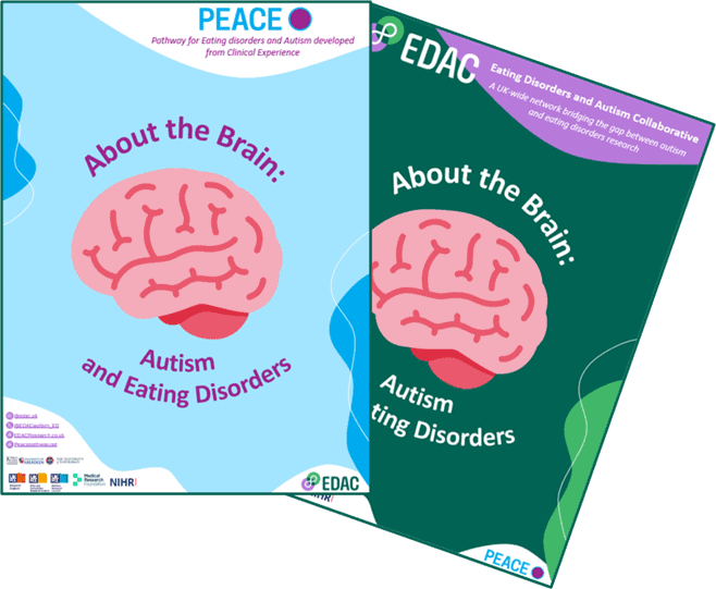
During this research exchange, I was able to foster strong relationships with researchers at KCL who are associated with both the Institute of Psychiatry, Psychology and Neuroscience (IOPPN), and the clinical-based Pathway for Eating disorders and Autism developed from Clinical Experience (PEACE) network. Collaborating with these researchers has paved the way for future collaborative projects and publications. This included the development of a patient and public involvement (PPI) booklet focused on neuroimaging in autism and eating disorders, titled “About the Brain”, located on both the PEACE and EDAC websites.
Specifically, I was able to work with researchers who created and developed the BEACON dataset, which provided valuable insights on the data that could be extracted for the purposes of a longitudinal imaging analysis. Out of the original 183 individuals used in our previous cross-sectional analysis from the first, 84 contained scans from TP1 and TP2. This data is currently being analysed using a longitudinal pre-processing pipeline, in which cross-sectional scans are computed at each timepoint, an average “base” is generated between time-points, and the scan is then re-processed using the “base” brain. Alongside KCL researchers, I developed two main approaches to analyse the dataset post pre-processing. One approach will focus on a basic longitudinal approach examining how T1-weighted brain structure changes during weight restoration. An additional secondary approach will focus on investigating myelin maps both cross-sectionally and longitudinally by determining T1-weighted / T2-weighted ratios. Alongside these two approaches, I was able to facilitate a collaboration between myself, a researcher at the University of Edinburgh and KCL. This intended collaboration will focus on examining measures of brain structure and obsessive-compulsive disorder collected within the BEACON dataset. Longitudinal pre-processing also has potential to strengthen previous machine learning approaches conducted in our initial cross-sectional study by incorporating the difference in brain morphology between TP1 and TP2 as a random forest regression model feature.
This research exchange has directly contributed to several anticipated academic outputs. Three publications are expected to arise from this collaboration, focusing on a standard longitudinal analysis of T1-weighted data, a cross-sectional and longitudinal investigation of myelin maps using T1-weighted / T2-weighted ratios, and a brief clinical report focusing on weight restoration across a 2-year period from the BEACON dataset. An additional publication focusing on OCD-related measures is expected to arise as a collaboration between myself, the University of Edinburgh and KCL. These outputs will not only contribute to the field of eating disorders, autism and neuroimaging, but have also already fostered national and interdisciplinary collaboration.
I would like to extend my sincere thanks to SINAPSE, who has provided me with the opportunity to work on pre-processing TP2 scans from the BEACON dataset. This exchange has been highly beneficial for both the University of Aberdeen and KCL. This collaboration granted me access to a dataset that includes a cohort not yet captured at the University of Aberdeen, while also maximising the value of a dataset collected by KCL. Beyond these two participating institutions, the exchange also generated new research ties between the University of Edinburgh, and strengthened existing ties between members of the Eating Disorders and Autism Collaborative (EDAC) network. On a personal level, this opportunity has been invaluable as Professor Kate Tchanturia has granted me permission to utilise and examine this dataset for future fellowship applications and foster my career progression.
tACS-EEG study on elderly participants with and without hearing loss
Funding Recipient: Dr Tanja Atanasova, Psychology, University of Dundee
Supervisor: Dr Anne Keitel, Psychology, University of Dundee
Dr Basil Preisig, University of Zurich
Partner Organisation: University of Zurich. Switzerland
Project Report: The SINAPSE funding provided me with an invaluable opportunity to engage in a two-week research exchange, which significantly contributed to my academic and professional development. The experience allowed me to collaborate with leading researchers, engage in interdisciplinary discussions, and gain hands-on exposure to novel methodologies. This report outlines the key activities undertaken during my exchange, the benefits to my research and career, and the broader impact on the SINAPSE Network.
Activities During the Exchange
Collaboration with Dr. Basil Preisig
One of the highlights of my exchange was meeting Dr. Basil Preisig, who graciously hosted me at his lab. We had in-depth discussions about his academic journey, his research focus, and his career trajectory. These conversations were particularly inspiring as they provided insights into the challenges and opportunities within academia, helping me better understand potential pathways for my own career. Dr. Preisig also shared valuable advice on navigating interdisciplinary research, which aligns with my interests.
Engagement with PhD Students and Lab Environment
The lab environment was dynamic and rich in diversity, with PhD students from varied academic backgrounds, including phonetics, neuroscience, psychology, and linguistics. Interacting with them was an eye-opening experience. We discussed differences in academic paths, research methodologies, and approaches to addressing complex scientific questions. These exchanges deepened my appreciation for interdisciplinary research and highlighted the potential for collaboration across fields.
During my time in the lab, I was introduced to several cutting-edge facilities, including transcranial alternating current stimulation (tACS) and electroencephalography (EEG) setups. Observing these in action enriched my understanding of advanced neuroimaging techniques and their applications in cognitive neuroscience and language research.
Participation in Internal Colloquia and Lab Meetings
I attended an internal colloquium led by Prof. Sabine Stoll on language acquisition from an evolutionary perspective. This session provided fascinating insights into how language evolves and is acquired, contributing to a broader understanding of cognitive and linguistic processes. The colloquium also fostered stimulating discussions among participants, encouraging me to think critically about my own research questions.
In addition, I participated in a lab meeting from Nathalie Giroud’s group, which focused on reading in adolescents with impaired hearing, examined through EEG studies. This session was particularly relevant to my work, as it demonstrated innovative methods for investigating neural processing in populations with sensory impairments. The discussion emphasized the importance of tailoring research designs to specific populations, a lesson I aim to incorporate into my future projects.
Presentation of My Research
As part of the exchange, I presented my current research to the lab. This presentation was an invaluable opportunity to receive feedback from a diverse group of experts. The feedback I received was constructive and thought-provoking, offering new perspectives and potential directions for my work. The discussion that followed helped me identify areas where I could refine my methodologies and explore new research questions. The supportive and collaborative atmosphere made this a particularly rewarding experience.
Hands-On Experience with tACS Recording
One of the most exciting aspects of my exchange was participating in my first tACS recording session, which lasted four hours. This hands-on experience provided practical insights into the complexities of setting up and conducting such studies. It also highlighted the importance of meticulous preparation and teamwork in achieving reliable results. This exposure has motivated me to explore the potential applications of tACS in my own research.
Exposure to Interdisciplinary Collaboration
Overall, the lab’s emphasis on interdisciplinary collaboration was an inspiring aspect of my exchange. The integration of phonetics, neuroscience, psychology, and linguistics within the same research environment demonstrated the value of approaching scientific questions from multiple perspectives. This experience reinforced the importance of fostering collaborations across disciplines to address complex research questions effectively.
Benefits to My Research
The exchange provided several direct benefits to my research:
- New Skills and Techniques: Hands-on exposure to tACS recording and advanced EEG methodologies has expanded my technical skillset, which will enhance the rigor of my future
- Insightful Feedback: The feedback received during my research presentation has refined my approach and introduced me to new perspectives on data analysis and interpretation.
- Broader Understanding: Discussions with researchers from diverse backgrounds have deepened my understanding of interdisciplinary approaches, inspiring me to incorporate more integrative methodologies into my work.
Benefits to My Career
The exchange has also had a profound impact on my career development:
- Networking Opportunities: Building connections with researchers, including Preisig and his colleagues, has opened avenues for potential future collaborations.
- Professional Growth: The experience of presenting my work and engaging in high-level discussions has boosted my confidence and professional communication skills.
- Career Inspiration: Learning about the career paths of established researchers has provided valuable insights into navigating academia and pursuing interdisciplinary research.
Broader Impact on the SINAPSE Network
This exchange has contributed to the SINAPSE Network by:
- Fostering Collaboration: My interactions with researchers at the host lab have laid the groundwork for potential future collaborations, which could lead to joint projects and
- Knowledge Sharing: The insights gained during my exchange will be shared with my home institution and peers within SINAPSE, enriching the network’s collective expertise.
- Strengthening Connections: The exchange has strengthened ties between SINAPSE and the host institution, paving the way for continued partnerships.
Reflections and Future Directions
This exchange has been an immensely rewarding experience, both personally and professionally. It has provided me with fresh perspectives on my research, introduced me to new methodologies, and expanded my academic network. The interdisciplinary nature of the host lab has inspired me to seek out collaborations that transcend traditional disciplinary boundaries.
Moving forward, I plan to:
- Incorporate the feedback received during my presentation into my ongoing
- Explore opportunities to apply brain stimulations in my
- Maintain and strengthen the connections established during this
Acknowledgments
I would like to express my heartfelt gratitude to SINAPSE for providing the funding that made this exchange possible. I am also deeply thankful to Dr. Basil Preisig and his team for their warm hospitality and invaluable support throughout my stay. This experience has been transformative, and I am confident it will have a lasting impact on my research and career.
Conclusion
The SINAPSE-funded research exchange has been a pivotal experience in my academic journey. It has provided me with unique opportunities for professional growth, enriched my research with new skills and perspectives, and strengthened the collaborative ties within the SINAPSE Network. I look forward to applying the knowledge and insights gained during this exchange to my future endeavours, contributing to the advancement of research within the SINAPSE community and beyond.